Description
| REF. NO. LENGTH
644.050 50 mm 644.055 55 mm 644.060 60 mm 644.065 65 mm 644.070 70 mm 644.075 75 mm 644.080 80 mm 644.085 85 mm |
REF. NO. LENGTH
4644.090 90 mm 644.095 95 mm 644.100 100 mm 644.105 105 mm 644.110 110 mm 644.115 115 mm 644.120 120 mm |
| Technical Specification
Thread Diameter 12.5 mm Thread Length 22.0 mm Shaft Diameter 8.0 mm |
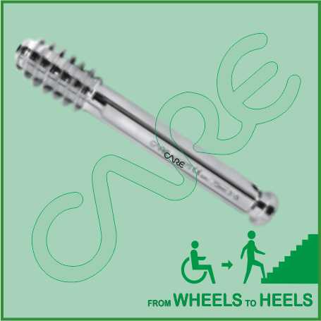
Hip Screw with Compression Screw 12.5mm is designed to provide strong and stable internal fixation of a variety of intertrochanteric, subtrochanteric and basilar neck fractures, with minimal soft tissue irritation.
Our Screws are made from finest quality medical grade material (Titanium and SS 316L) to ensure highest quality. Different sizes of Compression Screws are:
50mm, 55mm, 60mm, 65mm, 70mm, 75mm, 80mm, 85mm, 90mm, 95mm, 100mm, 105mm, 110mm, 115mm and 120mm.
Femoral Neck Fracture Hip Compression Screw 12.5mm
PRINCIPLES
Displaced transcervical and subcapital fractures are unstable. Their prognosis is by and large the same and they will be discussed as one group for the purpose of manner of reduction and choice of fixation, should internal fixation be chosen as the method of treatment.
If added rotational stability is desired in addition to the Hip Compression Screw, a cannulated screw is inserted above and parallel in both planes to the DHS. It must be parallel in order not to block the sliding property of the DHS implant.
Positioning of the patient
The patient is positioned supine on the fracture table for closed reduction. The ipsilateral arm is elevated in a sling and the contralateral uninjured leg is placed on a leg holder.
If closed reduction is not successful the patient may be transferred to a conventional table for open reduction.
C-arm image intensifier control during surgery is a must.
Approaches for open reduction
For this procedure the following approaches may be used for open reduction:
- Anterolateral approach
- Iliofemoral approach
REDUCTION
Closed reduction
Reduction can usually be obtained with gentle traction and internal rotation of the fractured leg, carried out under image intensifier control. The reduction must be checked in both the AP and lateral view with an image intensifier.
Occasionally, anteroposterior pressure applied to the thigh may help to reduce retroversion.
If gentle closed reduction is unsuccessful, proceed to open reduction.
The reduction should restore anatomical alignment.
Open reduction
If closed reduction fails, an open reduction must be carried out. The reduction of the neck fracture is carried out under direct vision.
Once the capsule is opened up while applying traction the head is manipulated with hooks or K-wires, inserted to act as joy sticks until an anatomical reduction is achieved.
FIXATION WITH DHS
Technique of insertion
The first step is to position a guide wire on the neck and hammer it into the head. With the C-arm positioned to show the neck axis, slide the guide wire along the neck, parallel to its axis, and gently tap it into the head.
With the C-arm in the AP, make sure that the wire subtends the CCD (collum-center-diaphysis) angle of the neck. This will help you with the insertion of the guide wire for the Hip compression screw.
Insertion of the guide wire
Choose the correct aiming device according to the CCD angle of the neck. Check its position in the AP view with the image intensifier.
Insert the guide wire through the aiming device and advance it into the subchondral bone of the head, stopping 10 mm short of the joint.
In both the AP and lateral planes, the guide wire should be positioned along the axis of the neck and through the middle of the head, and advanced to within 5 mm of the subchondral bone.
Determination of the length of the Hip Compression screw
Determine the length of the Hip Compression screw with the help of the measuring device. Select a screw which is 10 mm shorter than the measured length.
Drilling
Adjust the cannulated triple reamer to the chosen length of the Hip Compression screw. Drill a hole for the DHS screw and the plate sleeve.
Hip Compression Screw insertion
The correct Hip Compression screw is mounted on the handle and inserted over the guide wire. By turning the handle it is advanced into the bone. Do not push forcefully or you may distract the fracture.
In young patients with hard bone it is best to use the tap to precut the thread for the hip compression screw. Otherwise the DHS screw may not advance, and you may actually displace the fracture by twisting the proximal fragment as you attempt to insert the screw.
When the hip compression screw has reached its final position (checked with the image intensifier: 10 mm short of the subchondral bone in the AP and lateral), the T-handle of the insertion piece should be parallel to the long axis of the bone to ensure the correct position of the plate.
Fixation of the DHS plate
Generally, a two-hole DHS plate with the preoperatively determined CCD angle will be chosen. Take the plate with the correct CCD angle, slide it over the guide wire, and mate it correctly with the screw. Then push it in over the screw and seat it home with the impactor.
Importance of screw position in intertrochanteric femoral fractures treated by Hip Compression Screw
Background
Tip-apex distance greater than 25 mm is accepted as a strong predictor of screw cut-out in patients with intertrochanteric femoral fracture treated by dynamic hip screw. The aim of this retrospective study was to evaluate the position of the screw in the femoral head and its effect on cut-out failure especially in patients with inconvenient tip-apex distance.
Patients and methods
Sixty-five patients (42 males, 23 females; mean age of 57.6 years) operated by dynamic hip screw for intertrochanteric femoral fractures were divided in two groups taking into consideration the tip-apex distance less (Group A; 14 patients) or more (Group B; 51 patients) than 25 mm. Patient’s age and gender, follow-up period, fracture type, degree of osteoporosis, reduction quality of the fracture, position of the screw in the femoral head, number of patients with cut-out failure and Harris hip score were compared.
Results
The average follow-up time was 41.7 months. The mean tip-apex distance was 17.14 mm in Group A and 36.67 mm in Group B. One (7.1%) patient in Group A and three (5.8%) patients in Group B had screw cut-out. Except the screw position, no statistical differences were observed between two groups with regards to study data’s. The screw was placed in femoral head more inferiorly (p = 0.045) on frontal and more posteriorly (p = 0.013) on sagital planes in Group B, while central placement of the screw was present in Group A. The common characteristic of three patients with screw cut-out in Group B was the position of the screw which was located in femoral head more superiorly and anteriorly after an acceptable fracture reduction.
Conclusions
Peripheral placement of the screw in femoral head increases tip-apex distance. However, posterior and inferior locations may help to support posteromedial cortex and calcar femoral in unstable intertrochanteric fractures and reduce the risk of cut-out failure.
 CARE IMPLANTS & INSTRUMENTS


